Confocal laser scanning microscopy allows for high resolution, multi-channel 3-dimensional imaging of fluorescently-labeled or reflective specimens. The advantage over conventional widefield light microscopy is that the optics of the confocal microscope remove scattered light and light originating from outside the focal plane of interest, thus generating a high contrast “optical section.” Three-dimensional image reconstruction of serial optical sections as well as quantitative measurements may be performed using the microscope software.
The facility houses 3 dedicated confocal laser scanning microscopes:
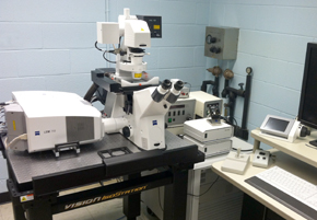
- a Zeiss LSM 880: 34 channel spectral system (32-channel array hybrid detector and two side PMT detectors, plus a transmitted light detector) configured around an AxioObserver (inverted) stand with a motorized XY stage. The system has six lasers: blue diode (405 nm and 440 nm), multi-line Argon (458, 488, 514 nm), yellow DPSS (561 nm), red HeNe (594 nm and 633 nm). The system is also equipped with an image scanning array detector (Airy) for high-resolution imaging. A stage incubator (which regulates temperature, CO2, and humidity) and objective heater are available to support live cell imaging.
- a Zeiss LSM 710: 34 channel spectral system (32-channel array detector (QUASAR) and two side PMT detectors, plus a transmitted light detector) configured around an AxioObserver (inverted) stand with a motorized XY stage. The system has five lasers: blue diode (405 nm), multi-line Argon (458, 488, 514 nm), green diode (561 nm), red HeNe (633 nm) and a 440 nm pulsed laser. The system is also equipped with a Becker & Hickl Fluorescence Lifetime Imaging system with 2 hybrid GaAsP detectors (for FRET-FLIM). The system is set up for FRET, FRAP and FLIM analysis. A low-profile PeCon stage incubator (which regulates regulates temperature, CO2, and humidity) and objective heater are available to support live cell imaging.
- a Zeiss LSM 700 configured around an AxioImager (upright) stand. The system has 4 solid state lasers (405 nm, 488 nm, 555 nm, and 635 nm). The scan head has 2 confocal PMTs, a variable secondary dichroic (VSD) beamsplitter and a transmitted light detector.
In addition to being able to tune to different emission wavelengths, both Zeiss array detectors allow for spectral scanning of fluorescence and emission “fingerprinting” as well as linear unmixing of signals from fluors with overlapping emission profiles. Both the LSM710 and the LSM 880 are also capable of FRET (fluorescence resonance energy transfer), FRAP (fluorescence recovery after photobleaching) imaging and FCS (fluorescence correlation spectroscopy).