Until recently, the best lateral resolution achievable by light microscopy has been governed by the theoretical diffraction limit of a microscope. In practical terms, under ideal conditions, this limit is approximately 200 nm lateral resolution (and approximately 500-700 nm axial resolution). Thus, in the XY plane, structures that are separated by less than 200 nm appear as single fused objects. Within cells, macro-molecular associations occur in spatial distances well below 200 nm.
Within the past 10 years, several different techniques (collectively known as “super-resolution microscopy”) have been developed that overcome the diffraction limit. The 3 most common of these techniques (and variations of these techniques) include:
- Point localization microscopy (e.g., Photo-activated Localization Microscopy (PALM) / Stochastic Optical Reconstruction Microscopy (STORM))
- Structured Illumination Microscopy (SIM)
- Stimulated Emission Depletion (STED) Microscopy
The SIM system housed in the MSR uses patterned illumination of a known spatial frequency (in multiple defined orientations) to improve resolution by extracting high spatial frequency information found in the fringes of the Moiré patterns formed by the interference of the sample (of an unknown pattern) with the illumination (known) pattern. The information derived from frequency space outside the observable region along with the observable data is processed to form the ‘super-resolution’ image, which has a lateral resolution approximately twice as good as that of conventional microscopy (i.e., one half the diffraction limit: 100 nm lateral; 270 nm axial).
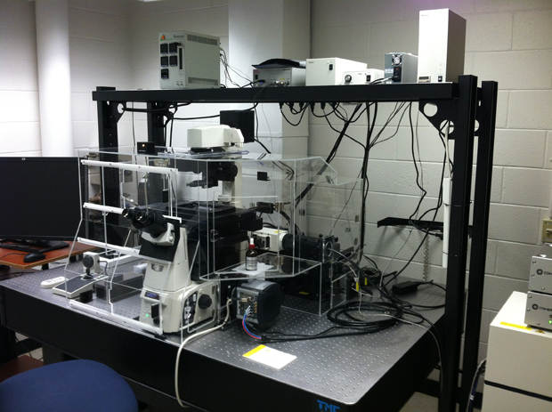
The facility houses two structured structured illumination microscopes:
- Zeiss Elyra 7 SIM/STORM: 10x, 25x multimmersion, 40x multimmersion, 63x SR oil (1.4 n.a., SIM) & 63x TIRF (1.49 n.a.) objective lenses. The system is equipped with two PCO edge sCMOS cameras (1280 x 1280 pixels) working in parallel.
- Nikon N-SIM: 10x, 20x, 40x, 60x water (1.27 n.a.) & 100x SR TIRF (1.49 n.a.) objective lenses. The system is configured with a Andor iXon DU-897E emCCD camera.
Both systems are equipped with:
- Multiple diffraction gratings, 4 lasers (405, 488, 561 & 640 nm)
- Hardware auto-focus module (laser-based automated focus drift compensation during live cell imaging)
- Motorized XY stage High-resolution piezo-driven Z stage
- Dedicated TIRF optics with independent laser input for TIRF (405, 488, 561 & 640 nm)
- Fluorescence & DIC optics
- Stage incubator (which regulates temperature, CO2 and humidity, to facilitate live cell imaging).
Both systems are capable of 3D-SIM (up to 20 µm deep), 2D-SIM (up to 3 µm deep), TIRF-SIM, and conventional multi-line TIRF and wide-field imaging.